
Page 14 - LabMedya - ENG - 07
P. 14
/labmedya
14 HEALTH AND LABORATORY MAGAZINE
AUTISM-ASSOCIATED
BRAIN DIFFERENCES CAN
BE OBSERVED IN PRENATAL
MRI SCANS IN THE WOMB
Study is the first to analyze
prenatal MRI scans of children
later diagnosed with autism.
A new study using prenatal cause challenges with the researchers used an ditions but not ASD.
brain scans revealed sig- communication, cognitive atlas-based automated
nificant differences in brain processing, emotional anatomical labeling meth- “Given that many genetic
structures at around 25 awareness and perception. od to segment the brain and environmental factors
weeks’ gestation between The causes of ASD are scans and then compared could affect the emer-
children who were later unknown but both genetic the segmented brain gence of ASD starting in
diagnosed with ASD and and environmental factors regions between the dif- the fetal stages, it is ideal
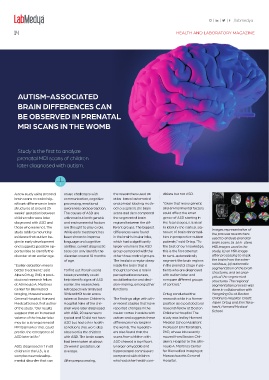
those who were not. The are thought to play a role. ferent groups. The biggest to identify the earliest sig- Images representative of
study adds to mounting While early treatment has differences were found nature of brain abnormal- the process researchers
evidence that autism be- been shown to improve in the brain’s insular lobe, ities in prospective autism used to analyze prenatal
gins in early development language and cognitive which had a significantly patients,” said Ortug. “To brain scans. (a-b) In-utero
and suggests possible op- abilities, current diagnostic larger volume in the ASD the best of our knowledge, MRI images used in the
portunities to identify the tools can only identify the group compared with the this is the first attempt study, (c) an MRI image
disorder at an earlier age. disorder around 18 months other three control groups. to semi-automatically after processing to mask
of age. The insula is a region deep segment the brain regions the brain from the exter-
“Earlier detection means inside the brain that is in the prenatal stage in pa- nal tissue, (d) automatic
better treatment,” said To find out if brain scans thought to have a role in tients who are diagnosed segmentation of the brain
structures, and (e) anal-
Alpen Ortug, PhD, a post- taken prenatally could perceptual awareness, with autism later and ysis of the segmented
doctoral research fellow help identify signs of ASD social behavior and deci- compare different groups structures. The regional
at Athinoula A. Martinos earlier, the researchers sion-making, among other of controls.” segmentation process was
Center for Biomedical retrospectively analyzed functions. done in collaboration with
Imaging, Massachusetts 39 fetal MRI brain scans Ortug conducted the Yangming Ou at Boston
General Hospital, Harvard taken at Boston Children’s The findings align with oth- research while in a former Children’s Hospital. Credit:
Medical School, first author Hospital. Nine of the chil- er recent studies that have position as a postdoctoral Alpen Ortug and Emi Taka-
of the study. “Our results dren were later diagnosed reported changes in the research fellow at Boston hashi, Harvard Medical
suggest that an increased with ASD, 20 were neuro- insular cortex in adults with Children’s Hospital. The School
volume of the insular lobe typical and 10 did not have autism and suggests these study was led by Harvard
may be a strong prenatal ASD but had other health differences may begin in Medical School Assistant
MRI biomarker that could conditions that were also the womb. The research- Professor Emi Takahashi,
predict the emergence of observed in the children ers also found that the PhD, whose lab recently
ASD later in life.” with ASD. The brain scans scans from children with moved from Boston Chil-
had been taken at about ASD showed a significant- dren’s Hospital to the Athi-
ASD, diagnosed in 1 in 68 25 weeks’ gestation, on ly larger amygdala and noula A. Martinos Center
children in the U.S., is a average. hippocampal commissure for Biomedical Imaging at
complex neurodevelop- compared with children Massachusetts General
mental disorder that can After preprocessing, who had other health con- Hospital.